

Currently, the gold standard for diagnostics of lung diseases is X-rays – such as, chest fluorography (radiography) and computed tomography (CT) of the lungs.
Compared to X-ray, ultrasound diagnostics has certain advantages, such as:
- absence of ionizing radiation;
- mobility/portability;
- availability.
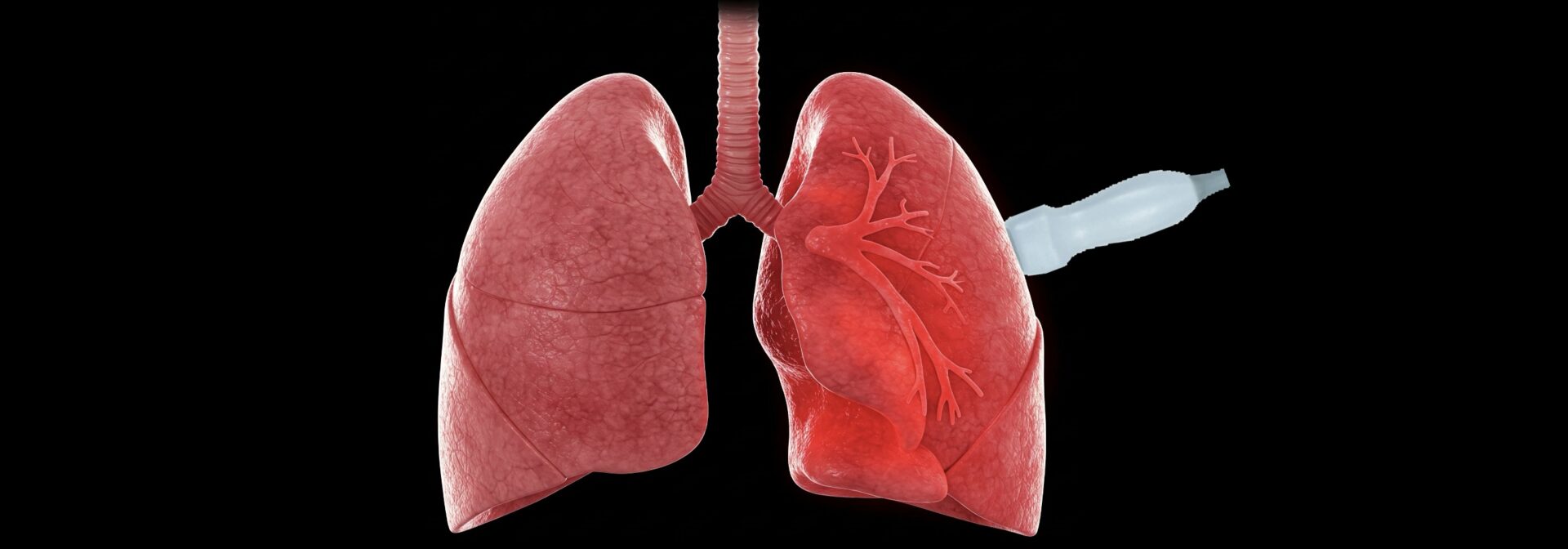
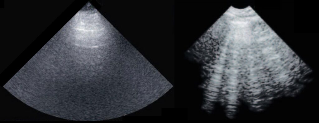
Interestingly, despite the fact that ultrasound virtually does not penetrate the lungs, reflecting from them due to a strong difference in the acoustic impedance of air and soft tissues, ultrasound images of diseased lungs (with pulmonary edema) differ significantly from those of healthy ones: vertical artifacts appear, called B-lines. However, the mechanism of B-line formation is not yet fully investigated, although its understanding is crucial for correct interpretation of lung ultrasound by physicians, as well as for the development of new methods of diagnostics.
LIMU tasks
- Investigation of physical mechanisms of B-line formation
- Development of new experimental setups
- Development of new theories of ultrasound lung diagnostics
Activity types
- experiments on tissue phantoms
- signal processing
- numerical experiment
- theory
Contacts
Details
- in a short video
- in the papers below
[1] Artifactual Lung Ultrasonography: It Is a Matter of Traps, Order, and Disorder / G. Soldati, A. Smargiassi, L. Demi, R. Inchingolo // Appl. Sci. 2020 — Vol. 10. — P. 1570. DOI: 10.3390/app10051570
[2] Physical Mechanisms Providing Clinical Information From Ultrasound Lung Images: Hypotheses and Early Confirmations / M. Demi, R. Prediletto, G. Soldati, L. Demi // IEEE Trans Ultrason Ferroelectr Freq Control. 2020. — Vol. 67, no. 3. — P. 612-623. DOI: 10.1109/TUFFC.2019.2949597