
The human brain is a complex object for ultrasound investigation and exposure due to the presence of cranial bones, which weaken and distort the ultrasound beam passing through them (i.e., introduce aberrations). Despite this, in recent years, the greatest clinical successes in the development of non-invasive ultrasound surgery (HIFU) are associated with brain surgeries to destroy tumors, treat essential tremor, symptoms of Parkinson’s disease, neurotic pain and other diseases.
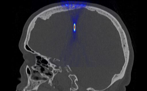
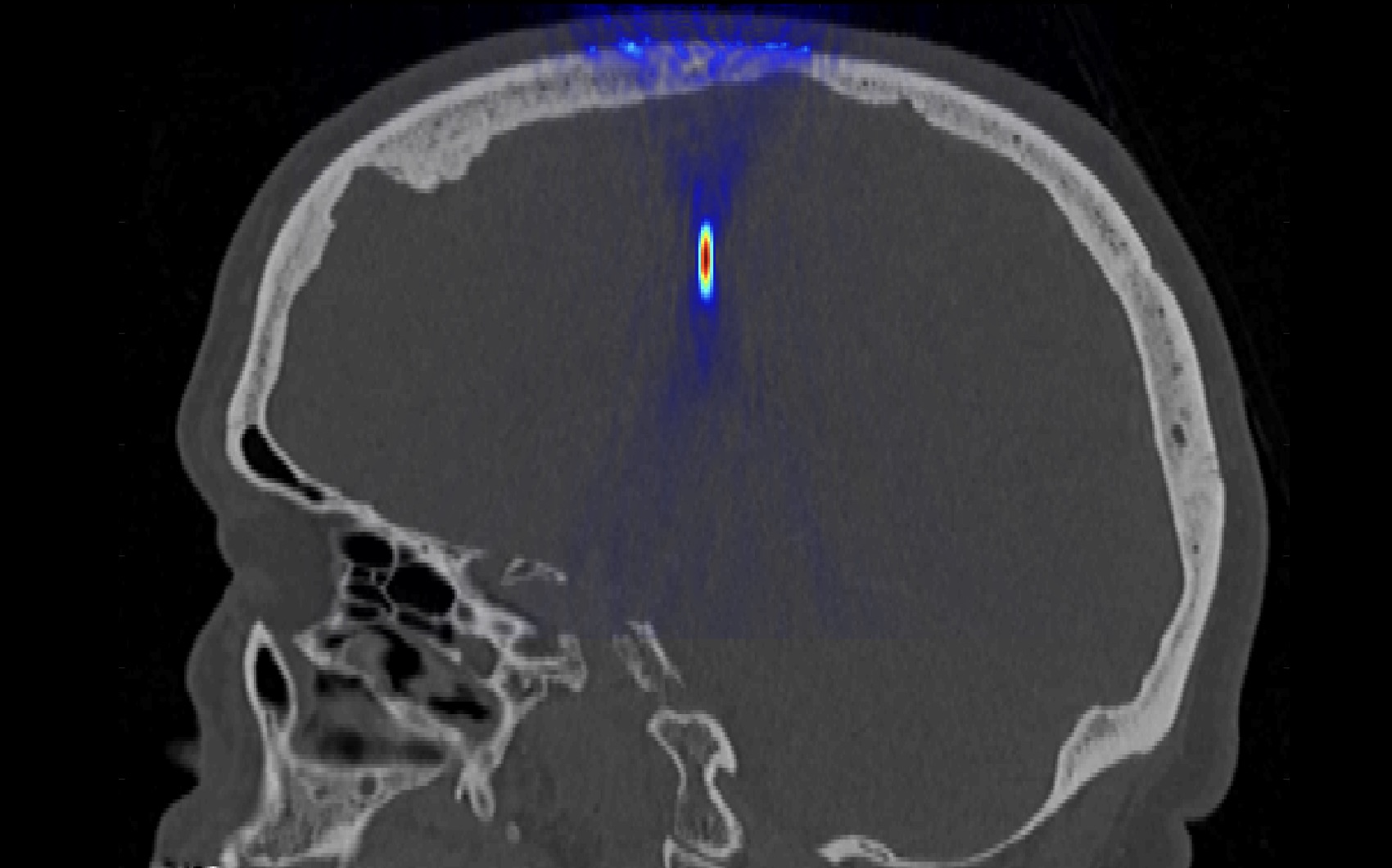
In the existing clinical HIFU system (Insightec, Israel), procedure planning is performed based on pre-operative computed tomography (CT) data, deriving an acoustic model of the head and modeling the ultrasound propagation and focusing in the target area of the brain with the ability to compensate for aberrations introduced by the non-uniform thickness of the cranial bones. At the same time, HIFU exposure itself is performed under MRI control. However, currently existing clinical systems only allow only exposure on central areas of the brain, although the causes of many diseases, including tumors, are often localized in other areas of the brain.
At LIMU, in cooperation with the University Clinic (MNOI) of Moscow State University:
- acoustic models of the human head with different properties are being developed using tomographic imaging data (CT and MRI);
- skull phantoms are being developed for physical and numerical experiments;
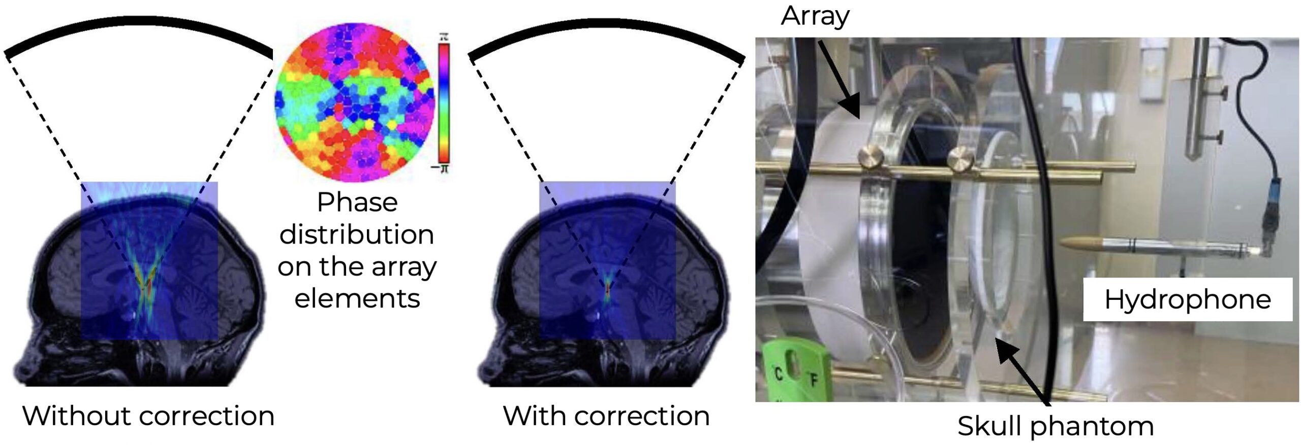
- a comparison is being made of the possibilities of compensating for aberrations using CT or MRI data;
- experiments are being performed to correct aberrations when focusing through already developed skull phantoms using a mosaic array developed at LIMU;
- various correction methods are being compared: ray-tracing methods based on geometric acoustics which are currently used in the clinical system, and new diffraction methods that provide high-quality correction in a wider range of focusing depths.
The ultimate goal is to produce an acoustic model of the human head to develop new methods of non-invasive ultrasound surgery using nonlinear waves and 3D printed skull phantoms.
Recently, there has also been increased interest in research into the possibilities of ultrasound brain diagnostics through an intact skull. In this case, compensation for aberrations introduced by the skull can be performed based on the information about its thickness profile obtained via computed tomography (CT) methods. A crucial task is to develop a fully ultrasound echo-pulse method for visualizing brain structures, based on thickness profile of the skull measured using ultrasound. We are conducting experiments on three-dimensional reconstruction of the external and internal surfaces of the skull using ultrasound, and developing methods for ultrasound visualization of scattering objects located behind the skull.
LIMU tasks
- Aberration correction when focusing ultrasound through the skull, using MRI and CT data
- Expansion of the area in the brain accessible for ultrasound exposure
- Development of methods for ultrasound diagnostics of the brain without the need for CT
Activity types
- numerical modeling
- experiments on skull phantoms
Contacts
- Alisa A. Krokhmal
- Daria D. Chupova
- Shamil A. Asfandiyarov
- Sergey A. Tsysar
- Vera A. Khokhlova
- Oleg A. Sapozhnikov
Details
[1] Ultrasound for the Brain: A Review of Physical and Engineering Principles, and Clinical Applications / W. Qiu, A. Bouakaz, E. E. Konofagou, H. Zheng // IEEE Trans Ultrason Ferroelectr Freq Control. 2021;68(1):6-20. DOI: 10.1109/TUFFC.2020.3019932
[2] The use of transcranial focused ultrasound in CNS diseases / M. V. Galkin // Burdenko’s Journal of Neurosurgery. 2016;80(2):108‑118. DOI: 10.17116/neiro2016802108-118
[3] Compensation for aberrations when focusing ultrasound through the skull based on CT and MRI data / D. D. Chupova, P. B. Rosnitskiy, O. V. Solontsov et al. // Acoustical Physics. — 2024. — Vol. 70, no. 2. — P. 288–298. DOI: 10.1134/s1063771024601651
[4] Compensation for aberrations of focused ultrasound beams in transcranial sonications of brain at different depths / D. D. Chupova, P. B. Rosnitskiy, L. R. Gavrilov, V. A. Khokhlova // Acoustical Physics. — 2022. — Vol. 68, no. 1. — P. 1–10. DOI: 10.1134/S1063771022010018
[5] A comparative study of experimental and simulated ultrasound beam propagation through cranial bones / A. Krokhmal, I. C.Simcock, B. E. Treeby, E. A. Martin // Physics in Medicine and Biology. — 2025. — Vol. 15, no. 70(2) — P. 025007. doi: 10.1088/1361-6560/ada19d
[6] Estimation of the thickness profile of a human skull phantom by ultrasound methods using a two-dimensional array / S. A. Asfandiyarov, P. B. Rosnitskiy, S. A. Tsysar et al. // Acoustical Physics. — 2023. — Vol. 69. — P. 112–118. DOI: 10.1134/S106377102270004X
[7] Simulation of nonlinear trans-skull focusing and formation of shocks in brain using a fully populated ultrasound array with aberration correction / P. B. Rosnitskiy, P. V. Yuldashev, O. A. Sapozhnikov et al. // Journal of the Acoustical Society of America. — 2019. — Vol. 146, no. 3. — P. 1786–1798. DOI: 10.1121/1.5126685
[8] Use of pulse-echo ultrasound imaging in transcranial diagnostics of brain structures / D. A. Sukhoruchkin, P. V. Yuldashev, S. A. Tsysar et al. // Bulletin of the Russian Academy of Sciences: Physics. — 2018. — Vol. 82, no. 5. — P. 507–511. DOI: 10.3103/S1062873818050283
[9] On the possibility of using multi-element phased arrays for shock-wave action on deep brain structures / P. Rosnitskiy, L. Gavrilov, P. Yuldashev et al. // Acoustical Physics. — 2017. — Vol. 63, no. 5. — P. 531–541. DOI: 10.1134/S1063771017050104
[10] A multi-element interstitial ultrasound applicator for the thermal therapy of brain tumors / M. Canney, F. Chavrier, S. Tsysar et al. // Journal of the Acoustical Society of America, 134 2 1647–1655. DOI: 10.1121/1.4812883